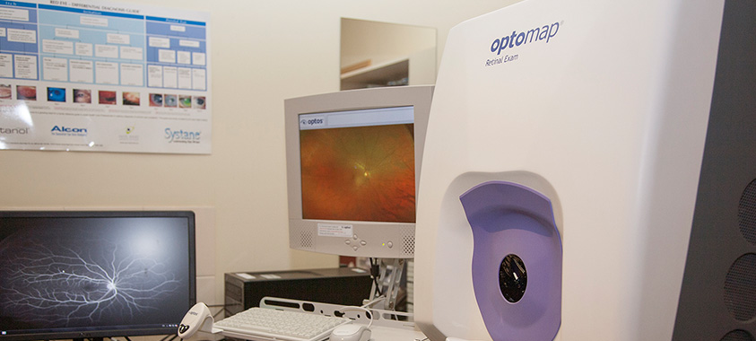
At Horsfalls, we are equipped with the latest state-of-the-art technology to ensure we are providing the best possible care and detecting any potential problems as early on as possible. This technology includes:
OCT (Optical Coherence Tomographer) – sometimes referred to as 3D retinal imaging, this machine is available in both Echuca and Kyabram. It is used to scan the optic nerve and/or macula to establish baseline values, detect eye disease (such as glaucoma and macula disease) and to monitor existing conditions. A scan takes just a couple of minutes and is painless. The resulting scan can be viewed immediately by the optometrist and patient and will show the microscopic layers of the retina.
OPTOS Optomap Retinal Camera – this camera was added to the Echuca practice in 2015. It takes a widescreen picture of the retina out to 200 degrees, is quick and painless and does not require dilated pupils. These wide angle photos aid in detection of peripheral retinal disease and are especially useful for patients with diabetes.
Medmont Visual Field Machines – these machines are in both our Echuca and Kyabram branches. The visual field test plots a map of the patient’s visual field, showing how widely and how easily they can pick up peripheral targets. Peripheral vision is important in mobility, driving and reading. Visual field testing is used in a wide variety of ocular and systemic conditions including glaucoma, stroke and retinitis pigmentosa. Sometimes a visual field test is required for renewal of a drivers licence.
Corneal Topographer – available in our Echuca practice, this machine is used to map the corneal surface for detection and quantification of astigmatism, and detection and monitoring of corneal disorders and diseases.
Anterior Eye Camera – this camera allows us to photograph anterior ocular lesions for monitoring.


